A new system has been developed in Russia for evaluating dental treatments using a training robot.

The article was published in the journal "Bulletin of PNIPU. Electrical Engineering, Information Technologies, Control Systems". The research was conducted with financial support from the Perm National Research Center "Rational Subsurface Use".
The project "Anthropomorphic Dental Simulator" is a training device for dental students, allowing them to safely practice essential procedures such as cavity treatment, tooth preparation for crowns, extraction, and root canal treatment. An integrated neural network utilizes cameras to evaluate the results of their work by processing the captured images.
Modern neural networks can identify numerous objects across different classes without the need for any additional schemes. Typically, a simple single-stage neural network is employed for detecting and classifying objects in photographs. For instance, it can accurately locate teeth in the simulator's jaw, even with constant changes in lighting and the shape of the object during treatment. However, if the analysis needs to focus on just a part of the object, such as a small filling, the task becomes more complex, increasing the likelihood of false positives. The neural network might mistakenly interpret reflections and irregularities inside the mouth as the desired openings in the tooth or completely overlook them.
Scientists from Perm Polytechnic University have developed a two-stage recognition scheme that analyzes photos to search for composite objects (individual teeth), extracts, normalizes their sizes, and examines each fragment separately to identify the sought-after small objects (fillings, openings).
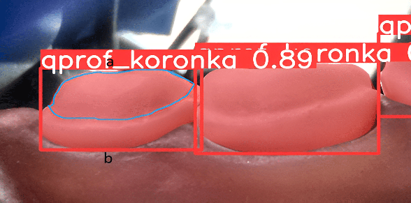
"In the first stage, the area of interest is identified, meaning the first neural network detects only the objects 'tooth' and 'tooth with a hole.' These are extracted and passed on to the second stage, where the openings in the teeth and their properties are recognized," explains Andrey Kokoulin, Associate Professor of the Department of Automation and Telemechanics at PNIPU, Ph.D. in Technical Sciences.
Preprocessing of photos is particularly crucial for determining the properties of small objects, as changes in them are harder to detect. It helps eliminate noise, enhance contrast and brightness, and improve clarity, making the image more informative. Because teeth have a color close to white, the outlines of the drilled openings are not easily visible. Additionally, reflections from the lighting necessary for the cameras' operation can interfere. The polytechnicians have also integrated a contrast enhancement program into the system that preserves local details and structures of the image, which is essential for accurately determining the boundaries of small objects in the image.
The shape of a tooth is a curve, and during the procedure, it is important to calculate the dimensions of its boundaries, the depth of openings, and the amount of enamel removed. To achieve this, scientists have developed a method for measuring complex-shaped objects, allowing calculations in three dimensions.
"The application of our two-stage system has increased accuracy by up to 92 percent and reduced the number of false positives by up to five percent. For each treatment option, the neural network can determine its quantitative parameters. For 'cavity' and 'canal' — the size of the cavity for the filling, for 'crown' — the thickness and uniformity of the layer removed from the sides and top of the tooth. It also assesses qualitative indicators — whether the treatment was performed correctly, if the tooth broke during extraction, and how even the walls are," shared Andrey Kokoulin.
The polytechnicians note that in the future, a mobile application could be created, allowing users to photograph a treated tooth (still without a filling or crown) and assess the quality of the treatment. The proposed analysis method can also be utilized anywhere that requires imaging of various composite structures and mechanisms with numerous details.
The system developed by PNIPU scientists based on neural networks significantly enhances the training of students on the dental simulator and contributes greatly to the advancement of modern technological medicine in Russia.